LI04Junction ValleyLarge Intestine Meridian4 of 18
No study in this index covers Myelitis Myatrophy Progressive
Every protocol below comes from a clinical text or a website. Nothing here has a published trial behind it, so no two independent sources have been shown to agree. How often that happens
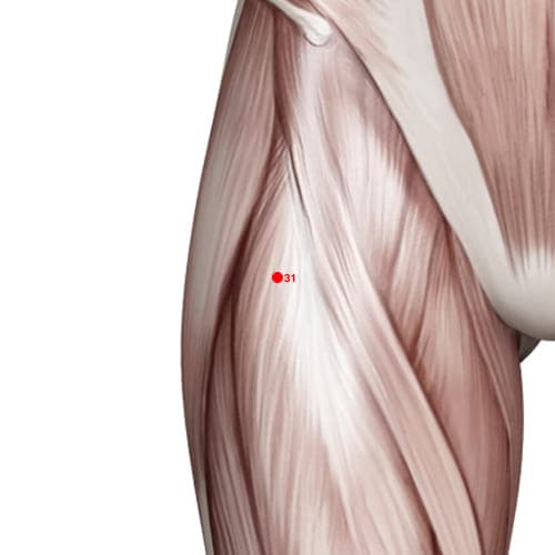
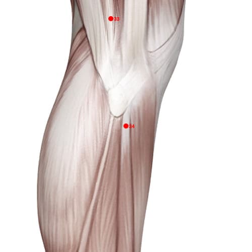
🎯 Location: On the anterior side of the thigh on the line connecting asis and laterosuperior corner of the patella on the level of the inferior pubis symphasis when thigh is fixed in the depression the lateral to the sartorius.
⚠️ Cautions: Caution with depth near femoral vessels.
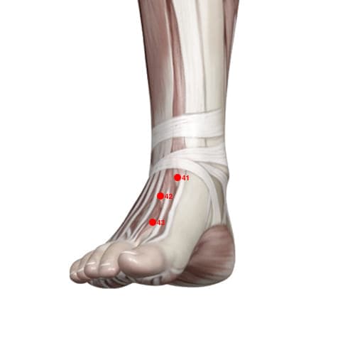
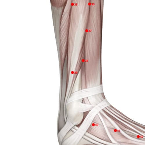
🎯 Location: On the dorsum of the foot at the midpoint of the transverse crease of the ankle joint in the depression the between tendons of the extensor digitorum longus and hallucis longus approximately at the level of the tip of the external malleolus.
⚠️ Cautions: Rule out fracture, dislocation and joint instability before needling, and do not needle into an acutely swollen joint or a haemarthrosis.
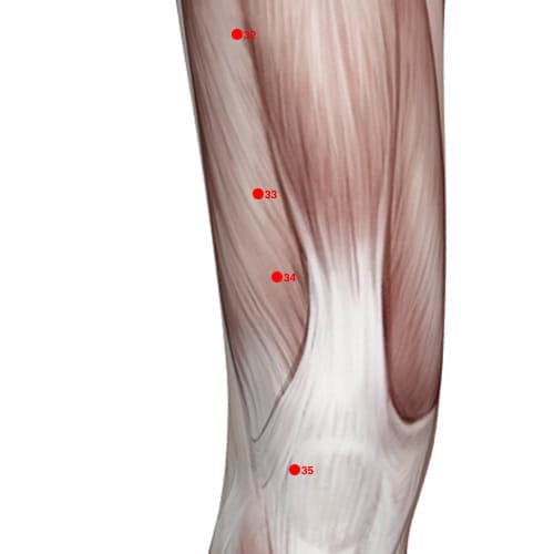
🎯 Location: When knee is flexed point is 2 thumb widths above the mediosuperior border of the patella on the bulge of the medial portion of the quadriceps femoris.
⚠️ Cautions: Anticoagulant therapy or a coagulopathy raises the risk of haematoma at the needling site – apply prolonged pressure on withdrawal. The precaution governs technique; bleeding disorders remain a treatable indication.
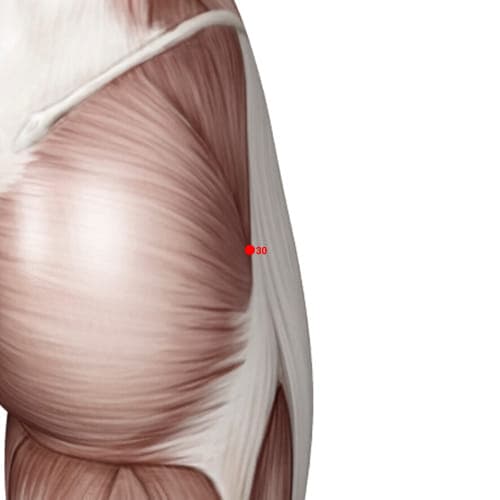
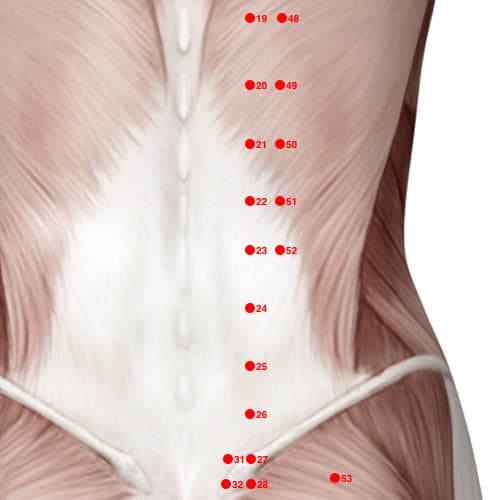
🎯 Location: At the junction of the lateral ⅓ and medial ⅔ of the distance between prominence of the greater trochanter and hiatus of the sacrum, GV02; when locating point put patient in the lateral recumbent position the with thigh flexed.
🎯 Location: On the anterior side of the thigh on the line connecting asis and laterosuperior corner of the patella on the level of the inferior pubis symphasis when thigh is fixed in the depression the lateral to the sartorius.
⚠️ Cautions: Caution with depth near femoral vessels.
🎯 Location: On the dorsum of the foot at the midpoint of the transverse crease of the ankle joint in the depression the between tendons of the extensor digitorum longus and hallucis longus approximately at the level of the tip of the external malleolus.
⚠️ Cautions: Rule out fracture, dislocation and joint instability before needling, and do not needle into an acutely swollen joint or a haemarthrosis.
🎯 Location: When knee is flexed point is 2 thumb widths above the mediosuperior border of the patella on the bulge of the medial portion of the quadriceps femoris.
⚠️ Cautions: Anticoagulant therapy or a coagulopathy raises the risk of haematoma at the needling site – apply prolonged pressure on withdrawal. The precaution governs technique; bleeding disorders remain a treatable indication.
🎯 Location: At the junction of the lateral ⅓ and medial ⅔ of the distance between prominence of the greater trochanter and hiatus of the sacrum, GV02; when locating point put patient in the lateral recumbent position the with thigh flexed.
🎯 Location: On the anterior side of the thigh on the line connecting asis and laterosuperior corner of the patella on the level of the inferior pubis symphasis when thigh is fixed in the depression the lateral to the sartorius.
⚠️ Cautions: Caution with depth near femoral vessels.
🎯 Location: On the dorsum of the foot at the midpoint of the transverse crease of the ankle joint in the depression the between tendons of the extensor digitorum longus and hallucis longus approximately at the level of the tip of the external malleolus.
⚠️ Cautions: Rule out fracture, dislocation and joint instability before needling, and do not needle into an acutely swollen joint or a haemarthrosis.
🎯 Location: When knee is flexed point is 2 thumb widths above the mediosuperior border of the patella on the bulge of the medial portion of the quadriceps femoris.
⚠️ Cautions: Anticoagulant therapy or a coagulopathy raises the risk of haematoma at the needling site – apply prolonged pressure on withdrawal. The precaution governs technique; bleeding disorders remain a treatable indication.
🎯 Location: At the junction of the lateral ⅓ and medial ⅔ of the distance between prominence of the greater trochanter and hiatus of the sacrum, GV02; when locating point put patient in the lateral recumbent position the with thigh flexed.
🎯 Location: On the anterior side of the thigh on the line connecting asis and laterosuperior corner of the patella on the level of the inferior pubis symphasis when thigh is fixed in the depression the lateral to the sartorius.
⚠️ Cautions: Caution with depth near femoral vessels.
🎯 Location: On the dorsum of the foot at the midpoint of the transverse crease of the ankle joint in the depression the between tendons of the extensor digitorum longus and hallucis longus approximately at the level of the tip of the external malleolus.
⚠️ Cautions: Rule out fracture, dislocation and joint instability before needling, and do not needle into an acutely swollen joint or a haemarthrosis.
🎯 Location: When knee is flexed point is 2 thumb widths above the mediosuperior border of the patella on the bulge of the medial portion of the quadriceps femoris.
⚠️ Cautions: Anticoagulant therapy or a coagulopathy raises the risk of haematoma at the needling site – apply prolonged pressure on withdrawal. The precaution governs technique; bleeding disorders remain a treatable indication.
🎯 Location: At the junction of the lateral ⅓ and medial ⅔ of the distance between prominence of the greater trochanter and hiatus of the sacrum, GV02; when locating point put patient in the lateral recumbent position the with thigh flexed.
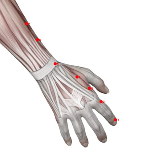
🎯 Location: On the dorsum of the hand between 1st & 2nd metacarpal bones approximately in the middle of the 2nd metacarpal bone on the radial side.
⚠️ Cautions: Traditionally forbidden in pregnancy – these are the points used to bring on labour at term. Controlled data show no excess of preterm birth or stillbirth (Carr 2015), but in pregnancy needle only on a clear obstetric indication.
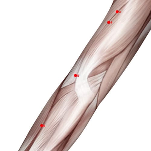
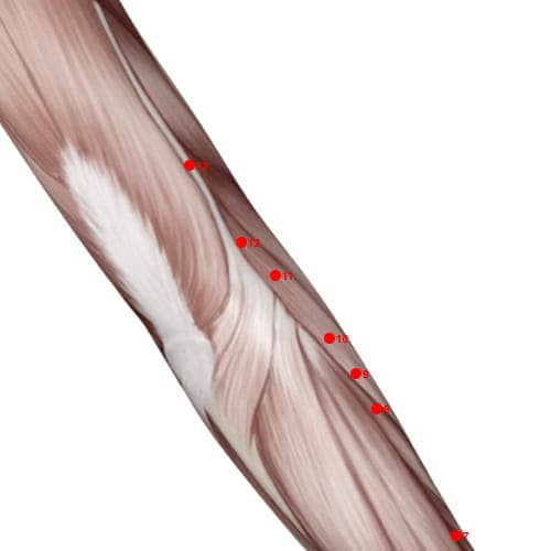
🎯 Location: When elbow is flexed point is in the depression the at the lateral end of the transverse cubital crease midway between LU05 and lateral epicondyle of the humerus.
⚠️ Cautions: Monitor blood pressure and avoid strong stimulation where hypertension is uncontrolled or unstable; hypertensive crisis is a medical emergency. Avoid excessive stimulation in weak patients.
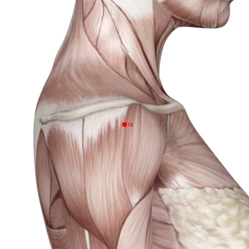
🎯 Location: Anteroinferior to the acromion on the upper portion of the deltoid muscle; when arm is in the full abduction the point is in the depression the appearing at the anterior border of the acromioclavicular joint.
⚠️ Cautions: Avoid deep perpendicular needling due to risk of pneumothorax; caution in shoulder injuries.
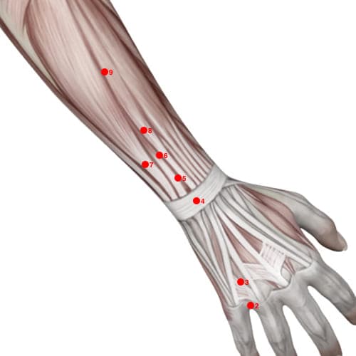
🎯 Location: 2 thumb widths proximal to the dorsal crease of the wrist on the line connecting TB04 and tip of the olecranon the between radius and ulna.
🎯 Location: On the dorsum of the hand between 1st & 2nd metacarpal bones approximately in the middle of the 2nd metacarpal bone on the radial side.
⚠️ Cautions: Traditionally forbidden in pregnancy – these are the points used to bring on labour at term. Controlled data show no excess of preterm birth or stillbirth (Carr 2015), but in pregnancy needle only on a clear obstetric indication.
🎯 Location: When elbow is flexed point is in the depression the at the lateral end of the transverse cubital crease midway between LU05 and lateral epicondyle of the humerus.
⚠️ Cautions: Monitor blood pressure and avoid strong stimulation where hypertension is uncontrolled or unstable; hypertensive crisis is a medical emergency. Avoid excessive stimulation in weak patients.
🎯 Location: Anteroinferior to the acromion on the upper portion of the deltoid muscle; when arm is in the full abduction the point is in the depression the appearing at the anterior border of the acromioclavicular joint.
⚠️ Cautions: Avoid deep perpendicular needling due to risk of pneumothorax; caution in shoulder injuries.
🎯 Location: 2 thumb widths proximal to the dorsal crease of the wrist on the line connecting TB04 and tip of the olecranon the between radius and ulna.